News & Events
Esophagogastroduodenoscopy (EGD)
Esophagogastroduodenoscopy (EGD)
What is an EGD?
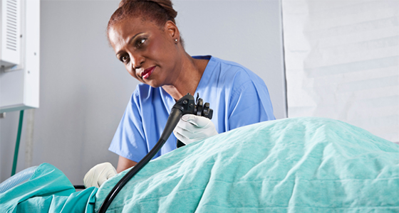
 An EGD, also known as esophagogastroduodenoscopy, is a procedure during which a panendoscope is used to examine the upper part of the gastrointestinal tract. A panendoscope is a long flexible tube that is thinner than most food you swallow. It is passed through the mouth and back of the throat into the upper digestive tract and allows the physician to examine the lining of the esophagus, stomach, and duodenum (the first portion of the small intestine).
Abnormalities suspected by X ray can be confirmed and others may be detected which are too small to be seen on X ray. If the doctor sees a suspicious area, he can pass an instrument through the endoscope and take a small piece of tissue (a biopsy) for examination in the laboratory. Biopsies are taken for many reasons and do not imply cancer.
Other instruments can also be passed through the endoscope without causing discomfort, including a small brush to wipe cells from the suspicious area for examination in the laboratory (a form of pap test or “cytology”) and a wire loop or snare to remove polyps (abnormal, usually benign, growths of tissue).
What preparation is required?
For the best possible examination, the stomach must be completely empty, so you should have nothing to eat or drink, including water, from 11 P.M. the evening before the examination or for at least 6 hours before its performance. Your doctor will be more specific about the time to begin fasting, depending on the time of day that your EGD is scheduled. Read detailed instructions about preparing for an EGD procedure or download a PDF version.
Be sure to let your doctor know if you are allergic to any drugs.
A companion must accompany you to the examination because you will be given medication to help you relax. It will make you drowsy, so you will need someone to take you home. You will not be allowed to drive after the procedure. Even though you may not feel tired, your judgment and reflexes may not be normal.
Please bring your X rays with you, as they may be important.
What should you expect during the procedure?
Your doctor will give you medication through a vein to make you relaxed and sleepy, and your throat may be sprayed with a local anesthetic. While you are in a comfortable position, the panendoscope is inserted into the mouth, and each part of the esophagus, stomach, and duodenum is examined.
The procedure is extremely well tolerated with little or no discomfort. Many patients even fall asleep during EGD.
The tube will not interfere with your breathing. Gagging is usually prevented by the medication.
What happens after the EGD?
You will be kept in the endoscopic area until most of the effects of the medication have worn off. Your throat may be a little sore for a couple of hours and you may feel bloated for a few minutes right after the procedure because of the air that was introduced to examine your stomach.
Are the any complications from EGD?
EGD is safe and is associated with very low risk when performed by physicians who have been specially trained and are experienced in this endoscopic procedure. Complications can occur but are rare.
One possible complication is perforation in which a tear through the wall of the esophagus or stomach may allow leakage of digestive fluids. This complication may be managed simply by aspirating the fluids until the opening seals, or may require surgery.
Localized irritation of the vein may occur at the site of the medication injection. A tender lump develops which may remain for several weeks to several months but goes away eventually.
Other risks include drug reactions and complications from unrelated diseases such as heart attack or stroke.
Death is extremely rare, but remains a remote possibility.
Why is EGD necessary?
Many problems of the upper digestive tract cannot be diagnosed by X ray. EGD may be helpful in the diagnosis of inflammation of the esophagus, stomach and duodenum (esophagitis, gastritis, duodenitis) and to identify the site of upper gastrointestinal bleeding.
EGD is more accurate than X ray in detecting gastric (stomach) and duodenal ulcers, especially when there is bleeding or scarring from a previous ulcer. EGD may detect early cancers too small to be seen on X ray and can confirm the diagnosis by biopsies and brushings.
EGD may also be needed for treatment, for example, for stretching narrowed areas of the esophagus or for the removal of polyps or swallowed objects. Active investigation is currently in progress on methods to control upper gastrointestinal bleeding through the panendoscope. Safe and effective endoscopic control of the bleeding could reduce the need for transfusions and surgery in these patients.
EGD is an extremely worthwhile and safe procedure which is very well tolerated, and is invaluable in the diagnosis and proper management of disorders of the upper digestive tract. The decision to perform this procedure was based upon assessment of your particular problem. If you have any questions about your need for an EGD, do not hesitate to speak to your doctor, who will also be happy to discuss the cost of the procedure, method of billing, and insurance coverage. Both of you share a common goal – your good health – and it can only be achieved through mutual trust, respect, and understanding.